TEM SPECIMEN PREPARATION
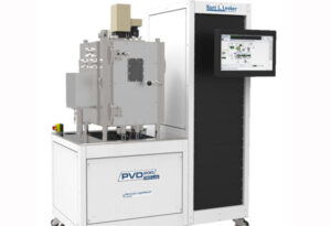
microPREP System
Enables fast, clean, and efficient preparation of samples for microstructure characterization and failure analysis. |
microPREP System - Advantages
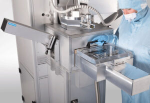
Introducing the microPREP™, a fast, clean, and efficient laser ablation system for optical, scanning electron microscopy (SEM), transmission electron microscopy (TEM), and focused ion beam (FIB) specimen preparation. It uses an ultra short laser pulse with micron level positional accuracy to rapidly produce samples with low structural damage. The microPREP system is ideal for sectioning advanced packages for subsequent isolation of through-silicon vias (TSV) and metal stack failures over a large length. Equally, the microPREP is the optimum tool for high volume applications that require large lamella TEM preparation. When used in combination with a FIB system, it not only shortens the FIB milling time, but significantly increases the overall throughput for large volume removal.microPREP system is best suited for:
- Fast sample preparation of metals and semiconductors, ceramics and compound materials
- Lamella preparation that requires positional accuracy of ±3 μm
- Increasing the workflow throughput of FIB and broad ion beam tools
- TEM, x-ray SEM (X-SEM), x-ray microscopy (XRM), advanced packaging tool (APT), and micromechanical testing
Benefits
- Shorter time to sample
- Analysis adopted sample geometry
- Minimizes risk of sample loss
- Recipe-driven GUI for sample geometries and materials
- Reduces FIB capacity requirements
- Enhances efficiency of existing (TEM) analysis tools
Applications
- High throughput and large area TEM lamella
- Fast slicing of centimeter size SEM cross sections
- Specific 3D shapes for x-ray microscopy
PIPS II System
Precision ion polishing system for precise centering, control, and reproducibility of your milling process. |
PIPS II System - Advantages
- X,Y stage permits alignment of argon beams to region of interest on the sample
- Improved collimated beam provides useable voltages as low as 100 volts for rapid and damage free preparation of FIB lamella
- Digital optical imaging with image storage and analysis in DigitalMicrograph® software
- 10" color touch screen for display and control of all PIPS™ II parameters
Dimple Grinder II
Fast and reliable mechanical method of pre-thinning to near electron transparency to greater reduce your ion milling time and uneven thinning.
Dimple Grinder II - Advantages
- Large transparent areas: Utilizes both the large wheel and flat wheel to preserve a large transparent area after processing
- Stronger specimens: Leaves a thick supporting rim to protect and strengthen the specimen after dimpling
- Direct preparation of TEM specimens: Produces a final thickness of <3 μm in dimpled specimens
- Accurate depth and thickness control: User-defined stop point and real-time display ensure you can produce the appropriate dimple depth and thickness
- Micro-positioning: Provides both orthogonal and intersect axes for more accurate positioning
Disc Punch System
Preferred method for cutting transmission electron microscope (TEM) discs from metals, alloys, and all ductile materials. |
Disc Punch System - Advantage
- Unique piston-rim design prevents overall compression and the cutting forces from extending into center region of specimen
- Exact fit of cutting and support pistons guarantees a sharp cut edge and easy removal of the punched disc
- Strong metal base ensures rigid and long term use without loss of precision
- Easy to see and remove punched area/disc simplifies collection and allows fast, single motion multiple punching
Disc Grinder System
Pre-thin and polish your sample to reduce ion milling times and improve quality |
Disc Grinder System - Advantage
- Lightweight grinder reduces maximum polishing pressure and specimen damage
- Achieve excellent control of specimen thickness with ultra-fine thread and pre-loading of the specimen drive screw
- Ensure uniformly thin specimens (5 µm accuracy) using large diameter polishing face with precise fit of the specimen mount
Ultrasonic Cutter
Cut your brittle materials beyond the 3 mm transmission electron microscope (TEM) disc to accurately cut hole or unique shapes. |
Ultrasonic Cutter - Advantage
- Quickly cut simple holes, unique shapes, or TEM discs from hard, brittle materials such as semiconductors, ceramics, and geological materials
- Cut materials ranging in thickness from <40 µm to 5 mm utilizing a piezo-electric crystal driving a tubular cutting tool in a fine grain, boron carbide slurry
- Minimize mechanical and thermal damage using manually tuned frequency driver to optimize cutting speed
- Reduce edge chipping and sample damage with spring-loaded platform and magnetically held table to prevent lateral movement
- Precisely center your site specific area with integrated stereo microscope and X,Y table
Cryoplunge 3 System
Semi-automated plunge freezing instrument. |
Cryoplunge 3 System - Advantage
Cryoplunge™ 3 system with GentleBlot™ technology is used for the preparation of frozen hydrated specimens for cryo-electron microscopy (cryo-EM).
- GentleBlot technology optimizes blotting force and pressure to provide gentle 1- and 2-sided blotting to minimize breakage of fragile specimen supports
- Humidified plunging chamber safeguards the specimen from drying during the blotting process
- Temperature controlled pot maintains the condensed ethane at its melting point
- Specially designed quick disconnect mechanism on plunge rod allows fast and easy attachment of the plunge tweezers to facilitate specimen handling
- Removable liquid nitrogen workstation provides a contamination free environment to protect the frozen hydrated specimen
Solarus Advanced Plasma Cleaning System
Advanced plasma cleaning system for removal of hydrocarbon contamination on TEM and SEM samples |
Solarus Advanced Plasma Cleaning System - Advantage
Solarus is a new generation plasma system engineered on new concepts and the latest technology resulting in better performance, simplified use, and consistent cleaning results. It is radically different from any plasma system available in the market and offers the most effective cleaning for removal of hydrocarbon contamination on transmission electron microscope (TEM) and scanning electron microscope (SEM) samples.
- Exclusive H2/O2 gas chemistry (patent pending)—this unique chemistry provides superior cleaning with less sputter damage of all samples including holey carbon films, and further enables samples to remain significantly cooler than if cleaned with the traditional Ar/O2 gas recipe
- Interactive touch screen interface offers seven pre-programmed recipes with optimized process parameters for greater consistency and repeatability
- Real time RF auto-match and variable RF power supply (10 – 65 W) ensure optimum plasma power and low loss under any cleaning conditions
- Mass flow controllers (MFC) provide accurate gas control for long term plasma stability and support up to three independent process gases H2, O2 and Ar
- Multi-functional chamber accepts two TEM holders through front ports while multiple SEM samples are loaded through the top opening lid
- Full time turbo drag pumping system enables overall faster cycle times—faster pump down (50 s vs. the competition's 3 min); faster venting (5 s vs. the competition's 6 min)
- Multi-language support includes operational instructions in English, German, French, Chinese, Korean, and Japanese
Cross Section Kit
Ideal for preparing cross sectional transmission electron microscopy (XTEM) samples of semiconductor devices, thin films of various substrates, and composite. |
Cross Section Kit - Advantage
- Quickly and easily prepare XTEM samples for TEM study of interfaces with greater consistency
- Used in conjunction with the ultrasonic cutter, cut rectangular areas of interest from bulk material to produce coated, stacked, and pressure-cured sections with minimum glue thickness (<1 µm)
- Suitable for preparing cylinder sections for dimpling prior to ion milling
TEM Specimen Holders
TEM In-Situ Software
In-Situ Explorer
Full in-situ control and data handling within Gatan Microscopy Suite software. |
In-Situ Explorer - Advantages
In-situ microscopy is a valuable tool to understand how real-world conditions and stimuli interact with and affect the structures, properties, and processes of a material. With the advent of more precise environment control and faster recording speeds, in-situ microscopy now generates some of the largest, information-rich datasets and imposes increasing demands on data capture, exploration, processing, and mining.
To address these rapid advancements, Gatan in partnership with DENSsolutions now introduces the In-Situ Explorer, the first software to centralize component control and data correlation from in-situ stimuli as well as high-resolution spatial and temporal imaging. Through a single software interface, In-Situ Explorer now offers a systematic and accurate way to correlate recorded images from Gatan IS cameras (OneView® and K2® IS) with the in-situ thermal data from the sample holder (DENSsolutions Wildfire, Lightning, and Climate systems) responsible for the observed specimen changes.
- Single control interface: Acquire, store, and analyze Gatan imaging and DENSsolutions holder data through Gatan Microscopy Suite® software
- Always know the sample temperature: Record actual temperature, set point, and heater power for every image
- Extract data based on time, temperature, and region of interest: Use the temperature history to explore and extract the precise data to be analyzed
TEM Holding Systems
Heating In-Situ Holders
Single and double tilt heating holders for direct observation of micro structural phase changes, nucleation, growth, and dissolution processes. |
Heating In-Situ Holders - Advantages
Heating in-situ holders – offered through partnership with DENSsolutions1
- The ultimate in sample stability at high temperatures
- Precise temperature control of specimen is provided using a MEMS microheater and true, 4-point, close loop control system
Model number | Max. temperature (°C) | Sample drift (nm/min at 800 °C) | Temperature control | Specimen positioning | Max. electrical feedthroughs |
---|---|---|---|---|---|
Wildfire | 800 (ST) 1300 (XT) | <0.5 | MEMS ST and XT 4-pt closed loop | α, β tilt | N/A |
Lightning (heating & electrical) | 800 (heating + bias) 1300 (heating) | <0.5 | α, β tilt | 4 | |
Climate (heating & gas) | 800 | <4 | α tilt | N/A |
Wildfire models
Wildfire S3 – FEI | Wildfire S5 – FEI | Wildfire D6 – FEI | Wildfire S3 – JEOL | Wildfire D6 – JEOL |
---|---|---|---|---|
±30° α tilt | ±70° α tilt | ±25° α and β tilt | ±20° α tilt | ±15° α and β tilt |
ST, XT, T, BioT | UT, ST, XT, T, BioT | ST, XT, T, BioT | HRP, HTP, CRP, HCP | UHP, HRP, HTP, CRP, HCP |
Wildfire Nano-Chips
Nano-Chip ST (RT - 800 °C) | Nano-Chip XT (RT - 1300 °C) | |
---|---|---|
Window materials:Silicon Nitride (window thickness 30 nm) | Window materials:Silicon Nitride (window thickness 20 nm) Carbon (window thickness 5 nm) Through-Hole |
BULK HEATING HOLDERS
- Precise temperature control of specimen holder tip is maintained using a bulk furnace and a water cooled specimen rod
- Heat loss from furnace to specimen tip is minimized using specialized ceramic furnace supports
Gatan model number | Maximum temperature2(°C) | Furnace | Specimen positioning | Specimen securing | Maximum electrical feedthroughs |
---|---|---|---|---|---|
628 | 1300 | Tantalum | α tilt | Hexring® mechanism | 6 |
628 | 900 | Inconel | α tilt | Hexring mechanism | 6 |
652 | 1000 | Tantalum | α, β tilt | Hexring mechanism | 2 – 63 |
652 | 850 | Inconel | α, β tilt | Hexring mechanism | 2 – 63 |
2For small pole piece gaps, the 628 maximum temperature is limited to 1000 °C for tantalum furnace and 700 °C for Inconel furnace, and the 652 maximum temperature is limited to 900°C for tantalum furnace and 700 °C for Inconel furnace
3Optional 2 – 6 for JEOL UHR, 2 maximum for other EMMs
Cooling In-Situ Holders
Optimized for low temperature observation of in-situ phase transitions. |
Cooling In-Situ Holders - Advantages
Transmission electron microscope (TEM) cooling holders are used in applications that require low temperature observation of in-situ phase transitions and to reduce contamination due to carbon migration.
- Reduce unwanted thermal effects in various analytical techniques and facilitates energy dispersive x-ray spectroscopy (EDS) and electron energy loss spectroscopy (EELS) analysis by reducing contamination and mass loss
- When configured for EDS applications, holder tip is designed to provide a clear path between sample and detector to reduce shadowing for EDS analysis
Cryo-Transfer Holders
Ideal for low-temperature transfer of frozen-hydrated specimens. |
Cryo-Transfer Holders - Advantages
Cryo-transfer holders are used in applications that require low-temperature transfer and observation of frozen-hydrated specimens for cryo-electron microscopy (cryo-EM).
- Cryo-workstation provides protected environment for loading specimen into holder
- Cryo-shield encapsulates frozen-hydrated specimen to protect it from contamination during atmospheric transfers from cryo-workstation to the transmission electron microscope (TEM)
Electrical In-Situ Holders
Measure electrical properties of samples with options for simultaneous temperature control and straining. |
Electrical In-Situ Holders - Advantages
Electrical in-situ holders – offered through partnership with DENSsolutions1
The Lightning in-situ TEM biasing & heating series provides you the power to obtain real-time information about your specimen under a controllable electrical and thermal environment. Investigate the next generation of nano-electronic materials and devices with the Lightning Series.
- The ultimate in sample stability at high temperatures
- Precise temperature control of specimen is provided using a MEMS microheater and true 4-point, closed-loop control system
- Multi-experiment Nano-Chip enables one device to support multiple samples and geometries
- Easy to use specialized optional FIB stub for simple sample alignment and placement
Gas In-Situ Holders
Characterized for gas-solid interactions up to atomic scale resolutions |
Gas In-Situ Holders - Advantages
Gas in-situ holders – offered through partnership with DENSsolutions1
The Climate in-situ TEM gas & heating series offers a high pressure gas environment at elevated temperature inside a sealable reactor – called the Nano-Reactor. Direct characterization of a solid-gas reaction at atomic resolutions can now be controlled and studied in real-time within your TEM. The Climate Series is a cost effective way of expanding and converting your high-end TEM from a static imaging tool into a fundamental real-world solid-gas research laboratory.
- The ultimate in sample stability at high temperatures
- Precise temperature control at all gas pressures with any gas mixture using a true 4-point, closed-loop control system
- Static and dynamic flow modes for wide-range of experiments
- Gas manifold with dynamic mixing reduces dead volume and provide quick adjustment to reaction gas
Straining In-Situ Holders
Quantify micro-structural changes in materials due to applied mechanical loading. |
Straining In-Situ Holders - Advantages
- Single push-button operation starts or stops the elongation process
- Gear reduction needed to provide small displacement rate required in TEM studies is achieved using a 2190:1 low-backlash, spur gear train followed by a 40:1 reduction precision worm and wheel drive
- Hexlok specimen securing mechanisms provides firm and secure clamping of the tensile specimen
Liquid In-Situ Holders
Discover biological and chemical phenomenon with samples in a fully hydrated environment. |
Liquid In-Situ Holders - Advantages
Liquid in-situ holders – offered through partnership with DENSsolutions1
The Ocean in-situ TEM liquid series empowers researchers to image materials and biological samples in a contained MEMS based cell – called the Nano-Cell. The Nano-Cell ensures the sample is fully hydrated and the liquid can be controlled in either a static or flowing condition. This Ocean Series opens up many new and exciting research fields, and transforms your TEM into a in-situ liquid characterization laboratory.
- High resolution to exceed the spatial resolution of optical microscopy for samples in liquid
- Modular design with removable tip prevents clogs and eliminates experimental cross-contamination
- Dual-orientation tip can be rotated by 180° to optimize for TEM or STEM imaging
- Native environment keeps the sample in a natural liquid state
- Controllable environment to adjust the type of liquid and flow rate around the sample
- Experimental safety with leak check tool enables known good liquid experiments to be loaded into the TEM
- Easy to use tip is designed for leak-free quick assembly without overpressure to Nano-Cell
Model number | Nano-Cell dimensions (mm) | Spacer thickness | Membrane thickness (nm) |
---|---|---|---|
Ocean | 2.00 x 2.60 | 200 nm, 5 µm | 50 |
Ocean models
Ocean S3+ FEI | Ocean S3+ JEOL |
---|---|
ST, XT, T, BioT | HRP, HTP, CRP, HCP |
Ocean Nano-Cell
1Limited availability to US, Japan, Canada, and India via distributor partnership with DENSsolutionsVacuum Transfer Holders
Preserve sample integrity with controlled transfer from an inert environment to the TEM. |
Vacuum Transfer Holders - Advantages
Transmission electron microscope (TEM) vacuum transfer holders are used in applications that require transfer of specimen from a preparation station (glove box or vacuum chamber) into TEM under vacuum or controlled atmospheric conditions.
- When configured for energy dispersive x-ray spectroscopy (EDS) applications, the beryllium cradle is designed to provide a clear path between the sample and the detector to reduce shadowing for EDS analysis
Model number | Operating temperature | Beryllium cradle | Specimen positioning | Specimen securing | Motorized control |
---|---|---|---|---|---|
648 | Ambient | √ | α, β tilt | Hexring®mechanism | β tilt |
VTST4006 | Ambient | α tilt | Clamping ring | ||
CHVT3007 | -160 to +50 °C | Optional | α tilt | Clamping ring1 |
1 Analytical configuration uses a Hexring mechanism.
Analytical Holders
Optimized for EDS analysis at ambient temperature. |
Analytical Holders - Advantages
TEM analytical holders are designed for basic imaging and analytical applications, such as electron diffraction and energy dispersive x-ray spectroscopy (EDS) analysis of crystalline TEM specimens.
- A beryllium specimen cradle minimizes unwanted x-ray signals
- Cut-outs in the specimen tip are matched to the TEM configuration and provide a clear path between the sample and detector to reduce shadowing for EDS analysis
- To facilitate quantitative x-ray analysis, non-rotating holder versions are equipped with a small Faraday cup located near the specimen cradle at the tip of the holder
- Electrical connections are available for the non-rotating versions of the holder
Model number | Beryllium cradle | Specimen positioning | Specimen securing | Motorized control | Faraday cup | Maximum electrical feedthroughs (optional) |
---|---|---|---|---|---|---|
643 | Standard | α tilt | Hexring®mechanism | Standard | 6 | |
646 | Standard | α, β Tilt | Hexring mechanism | β Tilt | Standard | 4 – 61 |
650 | Standard | α Tilt and rotation | Hexring mechanism | Rotation | No | 0 |
925 | Standard | α, β Tilt and rotation1 | Hexring mechanism | No | 0 |
Tomography Holders
High tilt and dual orientation holders for tomography applications. |
Tomography Holders - Advantages
Designed with low profile tips that allow up to 80° α tilt1 in small pole piece gaps providing a large, unobstructed field of view for tomographic data collection.
Model number | Minimum operating temperature | Specimen positioning1 | Specimen securing |
---|---|---|---|
916 | Ambient | α Tilt | Quickload™ clamping springs |
927 | Ambient | α Tilt, fixed 90° rotation | C-ring |
912 | Ambient | α Tilt | Specimen bonding to J- or C-shaped removable end tips |
914 | Less than -170 °C | α Tilt | Quickload clamping springs |
Multiple Specimen Holders
Accommodate multiple specimens for increased productivity. |
Multiple Specimen Holders - Advantages
Transmission electron microscope (TEM) multiple specimen holders provide increased productivity for applications that require comparable screening of multiple specimens.
- Operates at ambient temperature or cryo-temperature, depending on holder model
- Interchangeable cartridge design facilitates handling and storage of fragile specimens
- Opening on one side of FIBBEM™ cartridges allows access of the ion beam and reactive gasses to interact with the samples; cartridge can then be attached to the holder for viewing in the TEM
Model number | Specimen positioning | Specimen securing | Interchangeable cartridge | Operating temperature |
---|---|---|---|---|
677 | α Tilt | Hexring® mechanism | Yes1,2 | Ambient |
677.FIB | α Tilt | FIBBEM cartridge | Yes1 | Ambient |
910 | α Tilt | Splitring®mechanism | Yes3 | Less than -170 °C |
2 Small pole piece gap configuration does not have a removable cartridge.
3 Two specimen positions are available in holders for the Hitachi and Zeiss TEMs. Three specimen positions are available in holders for FEI, JEOL and Topcon TEMs.
Turbo Pumping Station
Vacuum Storage of holders and specimens, regeneration of cryo-specimen holders. |
Turbo Pumping Station - Advantages
The turbo pumping station is a completely self-contained, compact, benchtop pumping station designed to regenerate the sorb material in cooling and cryo-transfer holder dewars and for vacuum storage of transmission electron microscope (TEM) holders or samples.
- Incorporates the latest diaphragm pump and turbo drag pump technologies to produce a clean vacuum
- Pumping times from atmosphere to near base pressure are typically less than 2 minutes
- Work column mounted directly atop the turbo drag pump permits various pumping combinations for TEM holder and/or TEM sample storage
- Holder / sample storage capacity for four holders, four sample storage modules, or a combination of both
- Valves on the work column can be controlled to maintain vacuum when power is switched off making it possible to isolate the vacuum pumps from the work modules
TAC100 and TAC200 Series Anti-Contaminators
Minimize contamination in the vicinity of the specimen within the TEM |
TAC100 and TAC200 Series Anti-Contaminators - Advantages
The TAC100 series liquid nitrogen anti-contaminators act as miniature cryo-pumps to minimize contamination in the vicinity of specimens within the transmission electron microscope (TEM) optical column. The TAC200 liquid helium anti-contaminators are used with helium cooling holders.
- Blades of the TAC100 series anti-contaminators can be fully retracted or positioned near the tip of the TEM specimen holder using a three position bayonet collar
- Blades can be configured as single, double, or tubular
- Stainless steel flexible bellows allows movement of blades in and out of the specimen area of the objective lens without loss of vacuum
- Conduction rod and blades are electrically isolated from the TEM, which allows the blades to be linked to a separate touch alarm to warn the user in the event specimen holder is tilted too far
Model number | Cryogen | Minimum temperature | Standard gold plated copper blades diameter (mm) | Optional beryllium blades diameter (mm)1 | Blade positions | Hold time |
---|---|---|---|---|---|---|
TAC100 | Liquid nitrogen | Less than -180 °C | 1.0 | 0.5 | 3 | 6 |
TAC200 | Liquid helium | ~10 K | 1.0 | 0.5 | 3 | 4 |
TEM Imaging & Spectroscopy
EELS & EFTEM System
GIF Quantum Energy Filters
High throughput spectrometers to capture highly detailed data from your EELS and EFTEM experiments. |
GIF Quantum Energy Filter - Advantages
The GIF Quantum® filter represents the fourth and most advanced generation of post-column energy filters by Gatan. Redesigned from the ground up, the GIF Quantum filter combines advanced dodecapole-based electron optics with a blazing fast CCD camera system to yield an imaging filter that defines the new state-of-the-art in the capture of both highly detailed electron energy loss spectroscopy (EELS) and energy-filtered transmission electron microscope (EFTEM) data sets with maximum throughput.
- No compromise between EFTEM and EELS performance. Patented sensor readout architecture allows the same CCD sensor to be used interchangeably as a full-frame, high quality imaging device, a high speed live viewing device, and an ultra-fast spectroscopy device
- Aberration correction up to the fifth order allows the use of a 9 mm entrance aperture for EFTEM and 5 mm entrance aperture for energy loss spectroscopy at performance specifications superior to that of the third generation GIF Tridiem® filter
- Acquire images and spectra with unprecedented exposure control and dynamic range using integrated electrostatic shutter
- High speed (1000 spectra/s), dose-efficient scanning transmission electron microscope (STEM) EELS spectrum imaging for detail rich mapping
- Large field of view energy filtered imaging, mapping, and diffraction with narrow slit widths using 9 mm entrance aperture
- Improved collection efficiency for aberration corrected STEM EELS using 2.5 and 5 mm entrance aperatures
- Low noise imaging and high speed viewing from the same detector during dual-speed camera readout mode
- Broad range of operating modes and configurations from 60 – 300 kV
- Capture a broader range of edges (2000 eV range) in a single spectrum for simplified quantification
- Effortlessly measure precise energy shifts and apply advanced quantification routines for a new level of EELS analysis using DualEELS™ capability
- Increased confidence that imaging filter is operating at peak performance with advanced autotuning
- Outstanding energy resolution and very low image distortions using dodecapole-based optics
GIF Quantum SE system—Ideal for efficient filtered imaging and mapping while providing basic EELS support, and for tomography and beam sensitive samples when outfitted with a low dose scintillator. System comes standard with a 5 mm entrance aperture and the base performance of the Gatan US1000XP/FT camera system.
GIF Quantum ER system—System of choice for STEM EELS acquisition and fast EFTEM mapping from a broad range of electron sources. System comes standard with a 9 mm imaging aperture, STEM detector, electrostatic shutter, and the advanced modes of the US1000XP/FT camera system.
GIF Quantum ERS system—Highest energy resolution system available. Suitable only for monochromated electron sources. System comes with the same options as the GIF Quantum ER filter but also includes specialized electronics and components allowing it to break the 100 meV barrier.
For more information pertaining to EELS techniques, step-by-step instructions on analytical experiments, and access to downloadable spectra, please visit EELS.info, an educational site that is dedicated to supporting the EELS community.
Enfinium Spectrometers
Dedicated, hard-working spectrometers for your EELS experiments. |
Enfinium Spectrometers - Advantages
Enfinium™ spectrometers provide the technology of the GIF Quantum® energy filters in a dedicated spectrometer system. Redesigned from the ground up, the GIF Quantum filter combines advanced dodecapole-based electron optics with a blazing fast CCD camera system to yield an imaging filter that defines the new state-of-the-art in the capture of both highly detailed electron energy loss spectroscopy (EELS) and energy-filtered transmission electron microscope (EFTEM) data sets with maximum throughput.
Enfinium spectrometers provide the advanced EELS capability of GIF Quantum filters without the added expense of imaging and energy-filtered modes on the system. Two models are available to provide either basic EELS functionality or a fully enabled EELS system.
- Acquire spectra with unprecedented exposure control and dynamic range with the electrostatic shutter
- High speed, dose-efficient scanning transmission electron microscope (STEM) EELS spectrum imaging for detail rich mapping at 1000 spectra/s
- Simplified operation over most applications using fixed 3 mm entrance aperture
- Improved collection efficiency for aberration corrected STEM EELS using 2.5 and 5 mm entrance apertures
- 60 – 300 kV operation for a broad range of operating modes and configurations
- Capture a broader range of edges in a single spectrum for simplified quantification
- Measure precise energy shifts and apply advanced quantification routines for a new level of EELS analysis using DualEELS™ capability
- Improve spectral linearity for outstanding energy resolution with dodecapole-based optics
Enfinium SE—Workhorse EELS system combining simplified operation and spectral speed suitable for most basic EELS operations. System provides 3 mm fixed position EELS aperture, and base package provides 100 spectra/s rate in spectroscopy.
Enfinium ER—High performance EELS system incorporating the advanced features of the GIF Quantum ER filter in a dedicated spectrometer. System comes standard with 2.5 mm and 5 mm spectroscopy apertures, electrostatic shutter, and the advanced spectroscopy modes of the US1000XP/FT camera system.
ENFINIUM DEDICATED EELS SYSTEMS
Enfinum SE | Enfinum ER | |
---|---|---|
Electrostatic shutter | Optional | |
1000 spectra/s | Optional | |
Fixed 3 mm entrance aperture | √ | |
2.5 and 5 mm entrance apertures | √ | |
60 – 300 kV operation | √ | √ |
2000 eV EELS range | √ | |
DualEELS capability | Optional | Optional |
Dodecapole-based optics | √ | √ |
For more information pertaining to EELS techniques, step-by-step instructions on analytical experiments, and access to downloadable spectra, please visit EELS.info, an educational site that is dedicated to supporting the EELS community.
GIF Quantum LS Imaging Filter
Maximize your resolution and contrast for cryo-EM by combining low distortion GIF Quantum optics with a K2 Summit detector that offers a "frame rate 10x higher than other[s]" and produces "a DQE as high as 80%" |
GIF Quantum LS Imaging Filter - Advantages
The GIF Quantum® LS imaging filter provides fundamental insights into cellular organization and ultrastructure by combining high performance energy filtration with direct detection technology. Ideally suited for low dose imaging applications, researchers can maximize their cryo-electron microscopy (cryo-EM) and cryo-electron tomography capabilities to gain further insight into complex systems function and disease progression at the molecular level.
- Bringing together the low distortion optics of the GIF Quantum energy filter and the performance of the K2 Summit®
- Be confident while imaging smaller particles with a detective quantum efficiency (DQE) as high as 80% in conjunction with reasonably small exposure times
- Maintain high contrast with thick samples with the industry leading energy filter
- See more of your sample with a 4-fold increase in field of view
- Extend unattended throughput with fully automated multi-tilt and -region acquisition
Comparison between the resolution and molecule size for published single particle cryo-EM structures. The K2 Summit or Quantum LS have been employed in all the structures which define the high resolution frontier across a range of molecular sizes.
Digital Imaging Cameras
K2 IS Camera
Highest performance in-situ camera to resolve dynamic details in heating, catalysis, mechanical deformation, STEM diffraction, electrical testing, and chemical reaction experiments. |
K2 IS Camera - Advantages
Leveraging the speed, sensitivity, and radiation hardness of the proprietary K2 Summit®sensor, extend your research to the shortest time resolution with fully usable frames.
Counting and in-situ studies
- 14.2 Megapixel direct detection sensor reads out at 400 fps with each frame delivering a usable image
- Highest DQE maximizes signal-to-noise ratio
In-situ studies
- Full- or sub-area sampling at rates up to 1600 fps
- Direct data storage with >15 minutes capture time, regardless of frame rate
- Longest direct detector lifetime even when using highest dose rates
- Streamlines large dataset management for both novice and expert users
- Video buffer allows you to capture only the video you want; with post-event triggering and LookBack
- Never miss the start of an in-situ reaction again
Counting studies
- Retains full capabilities of K2 Summit modes (counting and super-resolution) for beam-sensitive materials for both materials science (zeolite, metal-organic framework (MOF)) and biological applications (cryo-tomography, SPA) structural characterization
K2 Direct Detection Cameras
Cryo-EM is being led by the K2 camera that offers a "frame rate 10x higher than other detector brands" and process "a DQE as high as 80%" |
K2 Direct Detection Camera - Advantages
“At present only the Gatan K2, when operated in counting mode, can produce a DQE(0) as high as 80% in conjunction with reasonably small exposure times. The K2 detector frame rate is about 10 times higher than that available with the two other detector brands.”– Subramaniam, S., Kuhlbrandt, W. & Henderson, R. (2016). IUCrJ 3, 3-7.
The K2 Summit® electron counting direct detection camera is simply the highest performance detector available for cryo-electron microscopy (cryo-EM). The unmatched frame rate of this direct detector allows electron counting modes to improve both contrast and resolution for large and small molecules alike.
- Only the K2 Summit can produce a DQE as high as 80%
- First to identify ground-breaking structural discoveries using cryo-EM
- K2® XP sensor provides the highest performance sensor for both resolution and contrast
- Superior detective quantum efficiency (DQE) means better science
- Image samples below 500 kDa at high resolution
- Reliably resolve differences with heterogeneous samples
- Higher quality cryo-tomography
- Super-resolution mode means 4x larger area in one image
Comparison between the resolution and molecule size for published single particle cryo-EM structures. The K2 Summit or Quantum LS have been employed in all the structures which define the high resolution frontier across a range of molecular sizes.
OneView Camera
Winner of the 2015 Microscopy Today Innovation Award. The ONE camera to capture 16 MP images and video in all your TEM applications. |
OneView Camera - Advantages
- Extends 4k x 4k camera leadership for imaging and in-situ data capture
- Always have a “live” experience with 25 fps at full 4k x 4k resolution, no compromise to resolution between viewing and recording images
- Guarantee optimal image quality with real-time drift correction and outlier removal using in-line data processing
- Detect single electrons with highest signal-to-noise ratio using the most sensitive scintillator and fiber optics available
- Extend the dynamic range beyond 16-bits, no beam stop required
- Increase productivity, even for novice users, with intuitive, built-in workflows to support and optimize recording modes
- Available as the primary camera for TEMs manufactured by FEI*, Hitachi High Technologies, and JEOL
In-situ option
- Flexibly trade-off resolution against frame rate—from 4096 x 4096 pixels at 25 fps to 512 x 512 pixels at 300 fps, always at 100% duty cycle
- Video buffer allows you to capture only the video you want; with post-event triggering with LookBack feature
- Never miss the start of an in-situ reaction again
- Tailor videos to your unique applications with powerful post-processing tools
Orius Cameras
Fiber-optically coupled CCD camera ideal for applications that require high sensitivity and pixel resolution. |
Orius Cameras - Advantages
- Most flexible range of applications including materials science, in-situ, biological, and diffraction covered by the Orius® family of cameras
- Fast frame rate with binning allows real-time setup and microscope optimization without viewing screen
- True transmission scintillator (Orius 833) offers best-in-class diffraction performance without worrying about damage
- Cameras are available both for bottom mount or 35 mm port locations for high resolution or high field of view applications
Model | 830 | 831 | 832 | 833 |
---|---|---|---|---|
Total megapixels | 4.2 | 7.1 | 10.7 | 4.2 |
Sensor pixel | 2048 x 2048 | 2672 x 2672 | 4008 x 2672 | 2048 x 2048 |
Pixel size (µm) | 7.4 | 9.0 | 9.0 | 7.4 |
Duty cycle (in view mode) | 100% | |||
Light coupling | Fiber optic | Lens-coupled | ||
True transmission scintillator | N/A | Yes | ||
Mounting position | On-axis or 35 mm port (retractable) | On-axis (retractable) | On-axis or 35 mm port (retractable) | 35 mm port (retractable) |
Digitization | 14-Bit | |||
Anti-blooming | On-chip (100x full well) | |||
Frame rate (fps, bin x4) | Up to 25 | Up to 14 | Up to 25 | |
Full well (CCD e-) | 40,000 | 50,000 | 40,000 | |
Non-linearity | <2% | |||
Exposure setting | 1 ms to 100 s | |||
TEM Shutter | Optional | |||
Computer interface | IEEE 1394b (Firewire 800) | |||
Regulatory compliance | EN 61326-1: 2006 | |||
IEC 61326-1: 2005 | ||||
EN 61000-3-2: 2006/A1; 2009/A2: 2009 | ||||
EN 61000-3-3: 2008 | ||||
IEC 61010-1: Third edition |
STEM Imaging Systems
STEMx System
A powerful tool that adds 4D STEM diffraction capabilities to your existing Gatan in-situ camera. |
STEMx System - Advantages
Introducing STEMx™, a 4D STEM diffraction system that collects data through hardware synchronization with Gatan’s in-situ cameras. By leveraging the high spatial and temporal resolution of the OneView® IS and K2® IS cameras, STEMx enables a wide variety of 4D STEM experiments, including strain mapping, indexing, orientation mapping, virtual apertures, and differential phase contrast.
- Leverage your existing Gatan in-situ camera for new 4D STEM applications
- Scan large sample areas with high diffraction space resolution
- Streamline your workflow for advanced applications
- Examine preliminary results in <10 min
Once a diffraction pattern is generated for each data point, a single, interactive Gatan Microscopy Suite® (GMS) 3 display enables you to view and manipulate all the information in a 4D STEM diffraction cube; including diffraction planes or individual diffraction patterns at each pixel location. STEMx is then able to process these large datasets within minutes using data size reduction methods that comprise of binning in four dimensions, sub-sampling in the spatial domain, or selecting a small region of interest. By consolidating both acquisition and analysis into GMS 3, not only can you quickly analyze and assess results at the microscope, but immediately decide whether to modify acquisition parameters or capture a new dataset.
Advanced STEM Detectors
High-angle annular dark field (HAADF), annular dark field (ADF) plus bright and dark field (BF/DF) detectors for STEM imaging optimized for electron energy loss spectroscopy (EELS). |
Advanced STEM Detectors - Advantages
For EELS mapping and scanning transmission electron microscopy (STEM) imaging, proper control of detector angles is critical in both data collection efficiency and its interpretation. The Advanced STEM detectors are optimized to work with EELS systems to provide seamless collection of the entire angular range of scattered electrons for an enhanced understanding of each sample’s chemical and compositional properties.
- Maximize electron collection using detectors optimized for EELS, diffraction contrast STEM and HAADF STEM applications
- Forward scattered electrons—EELS signal (0 to β), collection angle β regulated by STEM camera length
- Medium-angle electrons—Advanced BF/DF STEM detector (~β to 2x β)
- High-angle electrons—HAADF detector (~2x β to 7x β)
- Calibrate detector offset and system gain to allow absolute intensity measurements
- Low noise electronics and scintillator/photomultiplier tube (PMT) based design
- Detect single electrons at low fluxes
- Record high signal-to-noise ratio images at typical fluxes
- Optimize contrast in real-time using automated PMT gain selection
- Computer control of detector gain and insertion for remote operation
- One-click switching from dark field to bright field STEM mode (model 807)
- Ideal integration with GIF Quantum® and Enfinium™ EELS systems
- Compatible with Gatan cameras and DigiScan™ II system to upgrade existing STEM imaging installations
For more information pertaining to EELS techniques, step-by-step instructions on analytical experiments, and access to downloadable spectra, please visit EELS.info, an educational site that is dedicated to supporting the EELS community.
STEMPack Spectrum Imaging
A powerful method of obtaining detailed analytic data from a sample on an electron microscope equipped with scanning mode. |
STEMPack Spectrum Imaging - Advantages
The STEMPack™ system enables a powerful method of obtaining detailed analytic data from a sample on an electron microscope equipped with scanning mode. By acquiring multimodal data in scanning mode, a wealth of information from precise positions within a scanned image or line profile is acquired. This approach is the basis of a family of techniques known as spectrum imaging (SI).
Spectrum imaging systematically probes a defined specimen area to gather the maximum possible information in a fully automated process. The STEMPack system supports the acquisition of data from a broad range of detectors, including: electron energy loss (EELS), characteristic x-ray emission (EDS), cathodoluminescence emission (CL), absorbed / induced current (EBAC/EBIC), and electron diffraction. The resulting spectral data is then analyzed with powerful processing and visualization methods to reveal unique detail in the specimen. The combination of detailed spectral information with high spatial resolution is the most powerful aspect of electron microscopy and the STEMPack system can help you realize this power.
- Maximize data flexibility and artifact rejection with feature rich acquisition options
- Use a single application to analyze dense data sets as simply as a single spectrum; while directly cross reference data and results
- Acquire the data size and region you choose with fully configurable acquisition
- Jointly acquire virtually any signal produced in the electron microscope as part of a spectrum image
- Obtain high speed and dose-efficient data acquisition for detail rich mapping using direct hardware communication
- Both new users and seasoned practitioners can achieve outstanding results with simple, yet flexible user interface
For more information pertaining to EELS techniques, step-by-step instructions on analytical experiments, and access to downloadable spectra, please visit EELS.info, an educational site that is dedicated to supporting the EELS community.
STEM Cathodoluminescence
Vulcan Cathodoluminescence Detector
Gain insight into your sample's optical and electronic properties. |
Vulcan Cathodoluminescence Detector - Advantages
Characterize luminescence properties of materials at the nanoscale. The Vulcan™ detector is a cathodoluminescence (light) collection, detection, and analysis system for the (scanning) transmission electron microscope.
- Miniature mirrors enable installation in most mainstream TEM/STEM instruments
- High sensitivity allows easy analysis of luminescence from a wide range of samples
- Directly correlate sample morphology and microstructure with luminescence properties
- Measure electronic properties of luminescent materials at meV energy resolution including: band gap, and defect and quantum confinement effects in semiconductors
- Reveal surface plasmon resonance modes of nanoparticles in any STEM instrument
- Cryogenic cooling of the specimen increases luminescence efficiency, reduces damage caused by the electron beam, and reduces the line width of spectral features from many specimens
- Compatible with electron energy loss spectroscopy products from Gatan to allow correlation of absorption and emission processes
TEM Analysis
Gatan Microscopy Suite Software
Drives your digital cameras and surrounding components to support key applications including tomography, in-situ, spectrum and diffraction imaging, plus more. |
Gatan Microscopy Suite Software - Advantages
Gatan Microscopy Suite® (GMS) is the industry standard software for (scanning) transmission electron microscope experimental control and analysis. GMS 3 is completely revamped and uses a new, much simplified user interface. GMS 3 enables novice users to easily perform basic research applications, while it continues to provide the deep access and control highly experienced microscopists are used to and demand.
Selected benefits of GMS software:
- Technique-centric workflow: Guides you through experimental setup, execution and analysis
- Tabbed workspaces and data layout manager: Provide a user-friendly way to organize and manage data
- Dedicated view for live data: Clearly separates newly acquired data from prior experiments
- EELS made easy: New, powerful model-based EELS analysis routines provide higher quality results, easier
- Complemented by step-by-step instructions available on EELS.info, an educational site that is dedicated to supporting the EELS community
- DigitalMicrograph® scripting: Uncompromised full control for expert users
3D Reconstruction Module
Aligns and reconstructs your three-dimensional tilt series. |
3D Reconstruction Module - Advantages
The 3D Reconstruction software provides tools to precisely align the 2D projections collected by the 3D tomography software and generates a 3D volume from the aligned projections.
- Manual and automatic tilt series alignment with sub-pixel accuracy
- Assisted determination of tilt axis location
- Multiple reconstruction algorithms including weighted back projection (WBP) and the simultaneous iterative reconstruction technique (SIRT)
- Memory conserving version of the WBP and SIRT Algorithms
- Support for sub-volume reconstruction
- Live updates of volume during reconstruction
Comparison of reconstruction algorithms from simulated data. The left image shows the original object, a cube. Projections were generated from -60 to +60 degrees at 1 degree intervals. The middle image shows the cube reconstructed using weighted back projection. The right image shows the reconstruction using 20 iterations of the SIRT algorithm. The blue and red levels correspond to 30% and 60% of the maximum level in the reconstruction.
GPU Reconstruction Software
Reconstruct three-dimensional tilt series 100-times faster than conventional methods. |
GPU Reconstruction Software - Advantages
Acquire a quick tilt series, reconstruct the 3D volume, and optimize the acquisition conditions before acquiring a high quality dataset. Once you acquire the high quality data set, the 3D reconstruction takes minutes not hours.
GPU Reconstruction adds accelerated versions of the SIRT and SART algorithms to those listed on the reconstruction palette. The 3D Reconstruction software must be installed along with the accelerated GPU software.